M.E. Zaki,1 N.E. Hassan1 and S.A. El-Masry1
البيانات المرجعية لمحيط الرأس، في الأطفال والمراهقين المصريـِّين
مشيرة عرفات زكي، نيرة المرسي حسن، سحر عبد الرؤوف المصري
الخلاصـة: استخدمت البيانات المستمدة من دراسة مستعرضة شملت 26 8 27 من الأطفال الأصحاء في القاهرة، بمصر، لعمل مخططات منحنى نمو محيط الرأس واستنباط قيم مرجعية للنسبة بين محيط الرأس وطول الجسم في كلا الجنسين. وجمعت العينة من خلال مشروع منحنى النمو المعياري المصري الوطني للأطفال والمراهقين الذي أجري عام 2002. وتم الحصول على القيم الخاصة بالأتـراب (ذوي العمر الواحد) في كل شهر من الشهور الأربعة والعشرين الأولى من العمر، ثم للأتـراب في كل سنة من العمر حتى سن 18 عاماً. وقورنت هذه القيم مع تلك الخاصة بمجتمعات أخرى. وتُعَدُّ معايـير النمو التي تم استنباطُها مناسِبةً للاستخدام في برامج رصد النمو في جميع أنحاء مصر.
ABSTRACT: Data from a cross-sectional study of 27 826 healthy children in Cairo, Egypt, were used to construct standard growth charts of head circumference and reference values of relative head circumference to length/height for each sex. The sample was collected during the Egyptian Growth Curve Project for children and adolescents in 2002. Values were obtained for each month cohort for children aged 1–24 months, then for each year cohort until age 18 years. The values were compared with those of other populations. The constructed growth standards are suitable for growth monitoring programmes throughout Egypt.
Périmètre crânien – Données biométriques de référence pour les enfants et adolescents égyptiens
RÉSUMÉ: Les données collectées dans le cadre d’une étude transversale menée au Caire, en Égypte, et portant sur 27 826 enfants en bonne santé ont été utilisées pour construire les courbes de croissance normalisées du périmètre crânien et les valeurs de référence du périmètre crânien par rapport à la longueur/taille pour les deux sexes. L’échantillon de mesures a été collecté en 2002 dans le cadre du projet Egyptian Growth Curve Project for children and adolescents (projet pour l’établissement de courbes de croissance pour les enfants et adolescents égyptiens). Les valeurs ont été déterminées par cohorte mensuelle pour les enfants âgés de 1 à 24 mois, puis par cohorte annuelle jusqu’à l’âge de 18 ans. Ces valeurs ont été comparées à celles d’autres populations. Les normes de croissance ainsi construites sont pertinentes pour les programmes de surveillance de la croissance dans toute l’Égypte.
1Department of Bioanthropology, National Research Centre, Cairo, Egypt (Correspondence to S.A. El-Masry:
This e-mail address is being protected from spambots. You need JavaScript enabled to view it
).
Received: 23/09/05; accepted: 08/12/05
EMHJ, 2008, 14(1): 69-81
Introduction
Head circumference is one of the most significant findings in physical examination of children, especially in the evaluation of the development and early diagnosis of neurological disorders [1,2]. There is a rapid increase in head circumference and marked histological changes in the brain in early infancy [3,4]. Therefore, early recognition of deviations from normal growth is important.
Reference data for a large age range allow the follow-up of children and recognition of the catch-up growth in head circumference that can occur up to about 5 years, when the cranial sutures interlock [5–7]. Head circumference of school-aged children may prove a useful anthropometric tool in deciding early nutritional history [8,9]. Any significant reductions in head circumference found in malnourished children may have serious implications for their future performance and achievement [10].
In Egypt, there are no standard values for head circumference of children and adolescents that can be used as one of the standard charts for developmental evaluation. Our objective was to construct standard head circumference and relative head circumference references for each sex of Egyptian children and adolescents, from 1 month to 18 years of age, and compare our data with that of other populations.
Methods
Data were derived from head circumference measurements taken from Egyptian children and adolescents in a 2002 study in the Greater Cairo area to establish the Egyptian growth curve [11].
Sample
This was a cross-sectional study, including 27 826 healthy subjects (14 048 boys and 13 778 girls) grouped by age every month during the first 24 months of life, then every year until the age of 18 years for each sex separately.
Ideally, national growth curves would be based on a sample representing all areas of Egypt. The great majority of Egyptians live in the Nile river valley and a geographical gradient supposedly exists in the genetic constitution of Egyptians. However, the population of Cairo amounts to about one-fifth of the Egyptian population and most Cairo settlers have originated from all parts of Egypt. For this reason the Greater Cairo area represents much of the Egyptian population in a single site.
To establish a growth curve from which comparisons to normal can be made, it is necessary to obtain data from subjects with optimum growth opportunities. Therefore, the sample selection was confined to children from the higher socioeconomic groups who were assumed to provide optimum anthropometric data due to the low likelihood of their suffering malnutrition or overcrowding at home.
The sample was collected as part of the 2002 Egyptian National Standard Growth Curve Project [11] from children living in Greater Cairo: infants and preschool children attending 2 maternal and child welfare centres and 72 private kindergartens; and older children and adolescents attending 13 fee-paying primary, preparatory and secondary grade schools and 4 private sports clubs. All infants and children aged from 1 month to 18 years, who participated in the project were included in this study (i.e. selection was mainly based on age). Inclusion criteria included normal health in children who had a normal delivery after a normal pregnancy and with normal birth weight. Exclusion criteria included major genetic or organic diseases, which are known to affect normal growth patterns.
Data collection
The co-investigators in the project were professional doctors, well-trained in anthropometry. They provided training and participated in the data collection. Each team received 20 hours of classroom instructions over a 10-day period, which was followed by practical training for 4 hours per day for 2 weeks. The accuracy of the team measurements was evaluated during the training and during the field study by their respective supervisors. Quality control was accomplished on teams sequentially withdrawn from the field for re-evaluation. Intra-observer and inter-observer differences were periodically checked during fieldwork.
For each child in the sample, the following were performed:
- Questionnaire to parents, including personal data, socioeconomic data and the medical history of the child with special emphasis on chronic disease or long-term systemic treatment.
- Complete clinical examination to exclude organic or genetic disorders that might interfere with normal growth.
- Measurement of maximum head circumference using a non-stretchable tape and measurement of body length using an infantometer (children ≤ 3 years) or body height using a stadiometer (children 3+ years). Heights were measured to the nearest 0.1 cm, following the recommendations of the International Biological Program [12] and were recorded as the mean of 3 consecutive readings.
- The relative head circumference values were calculated as: [{head circumference (cm)/height or length (cm)} × 100].
Statistical analysis
Means and standard deviation (SD) of head circumference and relative head circumference were calculated for each age group and for each sex separately. The independent t-test was used in assessment of sex differences. Head circumferences at 3rd, 5th, 10th, 25th, 50th, 75th, 90th, 95th and 97th percentiles were determined for each sex and were graphically smoothed using a polynomial fitting equation. SPSS/PC software, version 8, was used for statistical analysis.
Results
Egyptian values
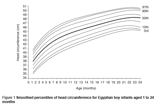
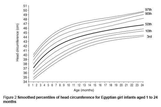
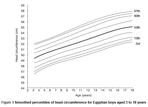
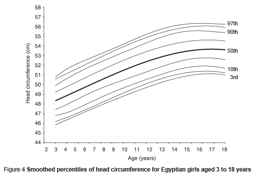
Means and SD of the head circumference values from 1 month to 18 years of age are presented in Table 1 for each sex separately. The means were significantly higher for boys than girls at each age (P < 0.001). They showed a progressive increase in head circumference values in all age intervals for both sexes. The highest increase was during the first 2 years of life with a large SD for both sexes: 6 cm during the first 6 months, 2 cm during the second half of the first year and 2 cm during the second year of life. The increase started to slow down (1.5 cm during the third year and about 0.5 cm at each year of life), until the age of 16 years for boys and 15 years for girls, with mean head circumference of 55 cm for boys and 54 cm for girls.
The mean height of boys was slightly greater than girls during the first 10 years of life; the differences were significant only at ages 4, 5 and 7 years. During ages 11 and 12 years, girls were significantly taller than boys (P < 0.05). Then boys became much taller than girls (P < 0.001) until the age of 18 years, where boys were 173 cm tall and girls were 159 cm tall.
Means and SD of the relative head circumference values are shown in Table 2 for ages 1 month to 18 years for each sex separately. During infancy, a significant difference was noticed during the first month of life, with boys having higher values. This difference disappeared up until the age of 11 months. However, during the second year of life, boys had significantly higher values than girls (P < 0.05), and this trend continued up to the age of 12 years (P 0.05). From the age of 13 to 18 years, girls had significantly higher values than boys (P 0.05). The smoothed means of the relative head circumference values showed a progressive decline in all age intervals for both sexes, until age 16 years for boys and 13 years for girls, when relative head circumference values started to be fixed (32 cm for boys and 34 cm for girls).
The smoothed percentiles of head circumference for Egyptian boys and girls aged from 1 to 24 months, and from 3 to 18 years, are presented in Figures 1, 2, 3 and 4.


International comparisons
The smoothed means of the head circumference values from the present data for the age range 1 to 24 months of life were compared with the data of Wikland et al. on infants in Sweden [13], Roche et al. on the population of the United States of America (USA) [14] and Attallah on infants in Saudi Arabia [15] (Figures 5 and 6). Comparing current data with that of Swedish and American infants, Egyptian boys and girls recorded lower values. However, compared with Saudi infants, Egyptian boys had higher values during the first 2 years of life. Egyptian girls recorded higher values during the first 6 months of life, then similar values until the age of 17 months, after which they had lower values at ages 18 and 24 months.
![Figure 5 Smoothed mean of HC of Egyptian boys aged from 1 to 24 months in comparison with American [16], Saudi Arabian [17] and Swedish [18] populations Figure 5 Smoothed mean of HC of Egyptian boys aged from 1 to 24 months in comparison with American [16], Saudi Arabian [17] and Swedish [18] populations](/images/stories/emhj/vol14/01/14-1-8-f5.png)
![Figure 6 Smoothed mean of HC of Egyptian girls aged from 1 to 24 months in comparison with American [16], Saudi Arabian [17] and Swedish [18] populations Figure 6 Smoothed mean of HC of Egyptian girls aged from 1 to 24 months in comparison with American [16], Saudi Arabian [17] and Swedish [18] populations](/images/stories/emhj/vol14/01/14-1-8-f6.png)
The data from 3 to 18 years of age were also compared with the American population data [14] and the Saudi Arabian population data [15] (Figures 7 and 8). Compared with American children, Egyptian boys and girls recorded lower values of head circumference (ranging between 1 to 2 cm for boys and about 1 cm for girls). Compared with Saudi children, Egyptian boys and girls had higher values, except at ages 6, 10 and 18 years for boys, and age 16 for girls, when they became equal. However at ages 17 and 18 years, Egyptian girls had lower values than Saudi or American girls.
![Figure 7 Smoothed mean of HC of Egyptian boys aged from 3 to 18 years in comparison with American [16] and Saudi Arabian [17] populations Figure 7 Smoothed mean of HC of Egyptian boys aged from 3 to 18 years in comparison with American [16] and Saudi Arabian [17] populations](/images/stories/emhj/vol14/01/14-1-8-f7.png)
![Figure 8 Smoothed mean of HC of Egyptian girls aged from 3 to 18 years in comparison with American [16] and Saudi Arabian [17] populations Figure 8 Smoothed mean of HC of Egyptian girls aged from 3 to 18 years in comparison with American [16] and Saudi Arabian [17] populations](/images/stories/emhj/vol14/01/14-1-8-f8.png)
Discussion
The growth supervision of children using growth curves is a widespread and useful tool in general paediatric practice [16]. Head circumference reference data is needed by health care professionals because this measurement is important in the assessment of growth and in screening for abnormal head size, which is related to the brain size and neurological status of infants and young children [17,18]. During infancy, the measurement and interpretation of head circumference has an important place in screening for conditions in which head size is abnormal, e.g. hydrocephalus and microcephaly. This measurement is less important during childhood and adolescence, but it is related to the extended observation of children whose head circumferences were unusual during infancy. After infancy, reference data can assist judgement of the normality of recorded measurements [14].
Since the head circumference standards show differences among races and generations, many researches have obtained normal values for their own populations, and recommend periodic re-evaluation of these standards [1,7,13,15,18]. The present head circumference data on Egyptian children and adolescents (aged from 1 month to 18 years) were collected in a cross-sectional study, which is a practical study design found widely acceptable to statisticians, as it is of high quality and quantity [19–21]. In addition, the head circumference measurement was recorded by well-trained and experienced professionals using standard and reliable equipment. Some specific features were included in the development of these updated reference values. The reference values are given not only for the measurement values, but also for the age-interpolated values and smoothed values. As children are rarely measured at exactly the same age in any growth study, this age interpolation and smoothing was felt to be necessary. This study provides the first reference standard of head circumference for Egyptian children aged 1 month to 18 years in a sample that was likely to represent all areas of Egypt.
The finding that head circumference was significantly larger in boys than in girls is in agreement with earlier reports [22,23] and with the general theory of the Y chromosome message [24].
There are possible international and inter-racial differences in head circumferences of children [25]. These could be observed by comparing the Egyptian data with that of Swedish infants [13], USA white children [14] and Saudi children [15]. Generally, comparing current data with that of Swedes and Americans, Egyptian boys and girls recorded lower values. However, compared with Saudi Arabians, Egyptians had higher values, except for girls during the second year of life and after 16 years of age. These differences can be explained in terms of the different population characteristics, genetic and nutritional, compared to the standards used in other populations. Tsuzaki et al. suspected that the relative head size would become similar in various populations if the nutrition and health of each population were optimal [25].
During infancy there was no significant difference between boys and girls in relative head circumference values. From the second year of life, boys had significantly higher values than girls and this trend continued up to the age of 12 years (except at age 10 years). Girls started to have significantly higher values than boys from the age of 14 years. This can be explained by the earlier pubertal spurt of girls, as boys are slightly taller than girls during the first 10 years of life whereas girls are significantly taller than boys during ages 11 and 12 years. Then boys become much taller than girls.
The present data will allow interpretation of head circumference values for adults, using the values of 16-year-old boys and 15-year-old girls as reference data, and for children older than 36 months, that could lead to the recognition of interfamilial clustering of unusual values.
References
- Karabiber H et al. Head circumference measurement of urban children aged between 6 and 12 in Malatya, Turkey. Brain & development, 2001, 23(8):801–4.
- Sanna E et al. The need of specific standards for head dimensions of urban Sardinian boys. Anthropologischer Anzeiger, 2003, 61(2):245–51.
- Rabinowicz T. The differentiate maturation of the human cerebral cortex. In: Falkner F. Tanner JM, eds. Human growth, 2nd ed. New York, Plenum Press, 1986, 2:385–410.
- Luo ZC et al. Growth in early life and its relation to pubertal growth. Epidemiology, 2003, 14(1):65–73.
- Roche AF. Possible catch-up growth of the brain in man. Acta medica auxologica, 1980, 12:165–79.
- Laron Z, Galatzer A. Effect of hGH on head circumference and IQ in isolated growth hormone deficiency. Early human development, 1981, 5:211–4.
- Al-Mazrou Y et al. Standardized national growth chart of 0–5-year-old Saudi children. Journal of tropical pediatrics, 2000, 46:212–8.
- Spurr GB, Reina JC, Barac-Nieto M. Marginal malnutrition in school aged Colombian boys: anthropometry and maturation. American journal of clinical nutrition, 1983, 37(1):119–32.
- Anzo M et al. The cross sectional head circumference growth curves for Japanese from birth to 18 years of age: the 1990 and 1992–1994 survey data. Annals of human biology, 2002, 29(4):373–88.
- Oyedeji GA et al. Head circumference of rural Nigerian children—the effect of malnutrition on brain growth. Central African journal of medicine, 1997, 43(9):264–8.
- Ghalli I et al. Egyptian growth curves 2002 for infants, children and adolescents. In: Proceedings of the 1st National Congress for Egyptian Growth Curves, Cairo University, 11 December 2003. Cairo, Department of Bioanthropology, National Research Centre, 2002.
- Tanner JM. Growth and physique: anthropometry. In: Weiner JS, Lourie JA, eds. Human biology: a guide to field methods. Philadelphia, F.A. Davis Company, 1969:2–42.
- Wikland KA et al. Swedish population-based longitudinal reference values from birth to 18 years of age for height, weight and head circumference. Acta paediatrica, 2002, 91(7):739–54.
- Roche AF et al. Head circumference reference data: birth to 18 years. Pediatrics, 1987, 79:706–12.
- Attallah NL. Patterns of growth of Saudi boys and girls from birth up to maturity in the Asir region: before the turn of the twentieth century. Saudi medical journal, 1994, 15:414–23.
- 16. Remontet L et al. Growth curves from birth to 6 years of age: growth in weight, height and cranial circumference according to sex. Archives of pediatrics, 1999, 6(5):520–9.
- Ross G, Lipper EG, Auld PAM. Growth achievement of very low birth weight premature children at school age. Journal of pediatrics, 1990, 117:307–9.
- Wellens R et al. Head circumference for Mexican American infants and young children from the Hispanic Health and Nutrition Examination Survey (HHANES 1982–1984): comparisons with whites and blacks from NHANES II (1976–1980). American journal of human biology, 1995, 7:255–63.
- Fomon J. Nutritional disorders of children: prevention, screening and follow up. Washington, DC, United States Department of Health, Education, and Welfare (DHEW Publication No. HAS 77-5104).
- National Center for Health Statistics. Growth charts. Health Resources Administration. Rockville, Maryland, US Department of Health, Education and Welfare, Public Health Service, 1976 (HRA 76-1120.25.3).
- Sullivan K. Anthro software computer package. Atlanta, Georgia, Nutrition Division, Centers for Disease Control, 1990.
- Brandt I. Dynamics of head circumference growth before and after term. In: Roberts DF, Thomson AM, eds. The biology of human fetal growth. London, Taylor & Francis, 1976:109–36.
- Karlberg P et al. Physical growth from birth to 16 years and longitudinal outcome of the study during the same age period. In: Karlberg P, Taranger J, eds. The somatic development of children in a Swedish urban community. Goteborg, Sweden. University of Goteborg, 1976:7–76.
- Ounsted C, Taylor DC. The Y chromosome message: a point of view. In: Ounsted C, Taylor DC, eds. Gender differences: their ontology and significance. Edinburgh, Churchill Livingstone, 1972:241–62.
- Tsuzaki S et al. The head circumference growth curve for Japanese children between 0–4 years of age: comparison with Caucasian children and correlation with stature. Annals of human biology, 1990, 17(4):297–303.


 Volume 31, number 5 May 2025
 WHO Bulletin
 Pan American Journal of Public Health
 The WHO South-East Asia Journal of Public Health (WHO SEAJPH)