H. Zedan,1 H.A. Abd El-Baset,2 A.A. Abd-Elsayed,3 M.F. El-Karn4 and H.R.H. Madkor 5
مستويات عامل نمو بطانة الأوعية في الأذيات الجلدية المرتبطة بالشدة السريرية (الإكلينيكية) لدى المرضى المصابين بالتهاب الجلد الأَرَجي بتماس الإسمنت
حاتم زيدان، هشام عبد الباسط، علاء عبد السيد، منى القرن، حافظ مدكور
الخلاصـة: يُعد التهاب الجلد الأَرَجي بتماس الإسمنت نمطاً متأخراً من التفاعلات المفرطة الحساسية التي قد يساهم فيها كل من غاما انترفيرون السيتوكينات وعامل نمو بطانة الأوعية في استمرار الاحمرار والوذمة. وقد قاس الباحثون هاتين المادتين لدى 32 من عمال البناء المصريـين المصابين بالتهاب الجلد الأَرَجي المزمن بتماس الإسمنت، ولدى 20 من الشواهد الأصحاء. واتضح للباحثين أن لدى هؤلاء المرضى مستويات زائدة زيادة يُعتد بها إحصائياً في كل من المصل وفي الجلد المصاب بالأذية، أكثر مما لدى الشواهد الأصحاء. ولاحظ الباحثون ترابطاً إيجابياً يُعتد به إحصائياً بين مستوى عامل نمو بطانة الأوعية وبين كل من مساحة الإكزيمة وحرز منسب الشدة لدى مرضى التهاب الجلد (r = 0.80). وهاتان المادتان قد تؤديان دوراً في إمراض التهاب الجلد الأَرَجي بتماس الإسمنت.
ABSTRACT Allergic contact dermatitis to cement is a delayed-type hypersensitivity reaction in which cytokines interferon-gamma (IFN-γ) and vascular endothelial growth factor (VEGF) may be involved in persisting erythema and oedema. VEGF and IFN-γ levels in serum and skin lesions were measured in 32 Egyptian building workers with chronic allergic contact dermatitis due to occupational exposure to cement and 20 healthy controls. Dermatitis patients had significantly higher levels of serum and lesional skin VEGF and IFN-γ than controls. A significant positive correlation was found between tissue VEGF and the eczema area and severity index (EASI) score in dermatitis patients (r = 0.86). VEGF and IFN-γ may play a role in the pathogenesis of cement allergic contact dermatitis.
Corrélation entre le taux de facteur de croissance de l’endothélium vasculaire des lésions cutanées et la sévérité clinique chez les patients souffrant de dermite de contact allergique due au ciment
RÉSUMÉ La dermite de contact allergique due au ciment est une réaction d’hypersensibilité retardée dans laquelle les cytokines interféron gamma (IFN- γ) et le facteur de croissance de l’endothélium vasculaire (ou VEGF pour vascular endothelial growth factor) peuvent être impliqués dans des érythèmes ou des œdèmes persistants. Les taux de VEGF et d’IFN- γ au niveau sérique et au niveau des lésions cutanées ont été mesurés chez 32 ouvriers du bâtiment égyptiens souffrant de dermite de contact allergique chronique due à une exposition professionnelle au ciment et chez un groupe témoin de 20 sujets sains. Les patients avec dermite présentaient des taux de VEGF et d’IFN- γ au niveau sérique et au niveau des lésions cutanées nettement plus élevés que le groupe témoin. Une étroite corrélation positive a été établie entre le taux de VEGF dans les tissus et le score de la surface eczémateuse et de l’indice de gravité (ou EASI pour eczema area and severity index) chez les patients atteints de dermite (r = 0,86). Le VEGF et l’IFN- γ peuvent jouer un rôle dans la pathogenèse de la dermite de contact allergique due au ciment.
1Department of Dermatology and Andrology; 2Department of Clinical Pathology; 3Department of Public Health and Community Medicine; 4Department of Physiology, Faculty of Medicine, University of Assiut, Assiut, Egypt (Correspondence to A.A. Abd-Elsayed:
5Department of Biochemistry, Faculty of Pharmacy, Al-Azhar University (Assiut Branch), Assiut, Egypt.
Received: 13/01/08; accepted: 27/03/08
EMHJ, 2010, 16(4):420-424
Introduction
Eczema and contact dermatitis (both irritant and allergic) account for 85%–90% of all occupational skin diseases in the developed countries [1]. Occupational allergic contact dermatitis is more prevalent than irritant contact dermatitis [2]. In developing countries such as Egypt, construction workers are frequently exposed to wet cement during their work in mixing, pouring and spreading the concrete or various cement mixtures due to little mechanization of the construction work. Hexavalent chromium is the most important allergen in patients with cement dermatitis [3,4].
Allergic contact dermatitis is mediated by a delayed-type hypersensitivity reaction. Previous experimental studies revealed that skin exposure to contact allergens induced a selective type 1 T-helper (Th1) cell response with increased production of the cytokine interferon-gamma (IFN-γ). Th1 cells play an important role in the acquisition of sensitization and the elicitation of contact allergic reactions [5–7]. Other mechanisms beyond this pathway have been implicated in contact hypersensitivity. Angiogenesis and its mediators are thought to play a critical role in this immune reaction [8,9]. Human vascular endothelial growth factor (VEGF) is a multifactorial cytokine that not only promotes angiogenesis but also enhances vascular permeability [10]. It has been reported that the expression of VEGF is increased in the lesional epidermis in inflammatory diseases such as psoriasis and contact eczema and in the serum of psoriatic arthritis patients [8,11,12]. VEGF might be involved in persisting erythema and oedema in eczematous skin [13].
The aim of the present study was to determine VEGF and IFN-γ levels in the serum and skin lesions of patients with cement allergic contact dermatitis to assess the involvement of these cytokines in the pathogenesis of this common type of eczema and to study the relation of their levels to the clinical severity of eczema.
Methods
Sample
This study included all building workers with chronic allergic contact dermatitis due to occupational exposure to cement (n = 32) who presented to University of Assiut hospital dermatology clinic in the period March 2006–February 2007. All the patients were males, age range was 18–52 years, mean age 33.5 [standard deviation (SD) 6.3] years. In addition, 20 age-matched healthy male volunteers, selected from visitors to patients in our department, were included as a control group.
All the patients with dermatitis had a long duration of cement exposure before development of the allergic contact dermatitis [range 6 months to 8 years, mean 3.5 (SD 1) years]. Repeated attacks or exacerbations were reported with every cement exposure, even minimal exposure, in all patients. The patients noted marked improvement with cessation of cement exposure. These features point to allergic contact dermatitis. The duration of dermatitis ranged from 1 to 5 years with a mean of 2 (SD 1.5) years. The affected sites were, in order of frequency: hands, forearms, feet, legs, arms, neck, thigh, trunk and face. All 32 patients showed variable degrees of lichenification and/or fissuring; 10 patients presented with acute exacerbations with weeping dermatitis, while 8 patients had subacute eczema with scaling and crusting in some lesions. The diagnosis was based mainly on clinical features and was confirmed using the thin-layer rapid use epicutaneous (TRUE) test which is a ready-to-use patch test system. All patients showed positive reactions to chromium (potassium dichromate patch) after application of the test to their back for 48 hours.
The severity of contact eczema in the patients was assessed using the eczema area and severity index (EASI). This includes an assessment of the surface area involved in 4 body regions: head and neck, upper limb, trunk and lower limbs (converted to a scale from 0–6). It also included assessment of erythema, infiltration/papulation, excoriation and lichenification, each on a scale of 0–3. EASI is a validated tool to objectively assess dermatitis severity with a maximum score of 72 [14]. In the present study, the score ranged from 4.5 to 39.1, with a mean of 26.7 (SD 10.1).
None of the patients included in the study had used any anti-allergic or anti-inflammatory therapy for at least 1 month. None of the patients had any other allergic conditions, dermatological disease, medical illness or malignancy. All patients and controls gave written informed consent.
Data collection
Blood samples (5 mL) were taken from patients and controls. Samples were allowed to clot for 30 min. at room temperature. Then samples were centrifuged for 10 min at 5000 rpm. Sera were separated and immediately stored at –70 °C until the time of assay.
Tissue samples were taken from both patients and controls. Punch biopsies (4 mm) were taken from the lesional skin in the patient group and from the inner aspect of the forearm near the elbow from the control group. Subcutaneous fat tissue was removed and the skin tissues were cut into small pieces on ice and were homogenized in 1/5 (w/v) 0.9% saline. Homogenates were centrifuged at 20 000 rpm for 15 min. Supernatants were separated and kept at –70 °C until the time of measurement. Care was taken to keep the temperature at –4 °C throughout the preparation of homogenates and supernatants.
Serum and tissue human VEGF levels were quantitatively determined in patients and controls by a solid phase sandwich enzyme-linked immunosorbent assay (ELISA), using a commercial immunoassay kit (R and D Systems, Minneapolis, USA). Serum and tissue human IFN-γ levels were measured in patients and controls by a commercial ELISA quantitative kit (Bender Med Systems, Vienna, Austria).
Statistical analysis
SPSS, version 15 was used for the data entry and analysis which included descriptive analysis, independent sample t-test, and Pearson correlation. The results were expressed as mean and SD. P-value
Results
Serum levels of VEGF ranged from 78.4 to 653.3 pg/mL, while lesional skin tissue levels of VEGF ranged between 38.0 and 57.8 pg/mg protein. Serum levels of IFN-γ ranged between 65 and 635 pg/mL and lesional skin tissue levels of IFN-γ ranged from 102.8 to 527.0 pg/mg protein. The mean values are shown in Table 1.
Patients with cement allergic contact dermatitis showed significantly elevated levels of serum and lesional skin tissue levels of VEGF compared with healthy controls (P = 0.005 and P = 0.001 respectively). They also had significantly higher serum and tissue IFN-γ levels compared with controls (P = 0.007 and P = 0.001 respectively) (Table 1). No significant positive or negative correlations were found between VEGF and IFN-γ levels in either serum or tissue.
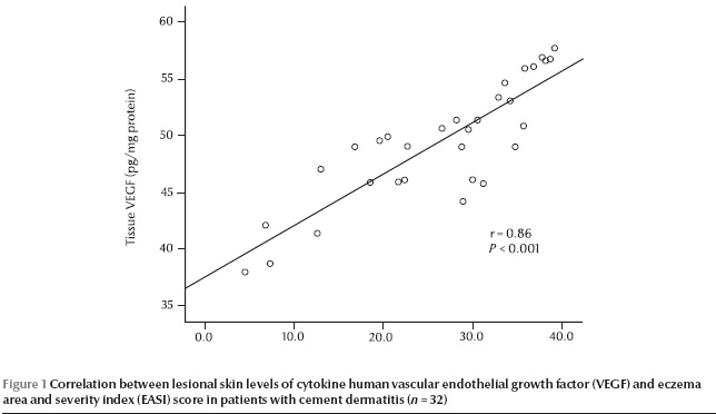
A significant positive correlation was demonstrated between lesional skin tissue levels of VEGF and the EASI score in patients with cement allergic contact dermatitis (r = 0.86, P Figure 1), while no significant correlations were detected between EASI score and other studied biochemical parameters.

Discussion
Allergic sensitization to occupational materials can result in serious morbidity, with time lost from work. Wet cement is an important cause of occupational skin diseases. Allergic cement eczema was found to have a greater extent of involvement than irritant cement eczema [15]. The pathogenesis of allergic contact dermatitis involves epidermal cells such as keratinocytes and Langerhans cells, fibroblasts of dermis and endothelial cells, as well as invading leukocytes interacting with each other under the control of a network of cytokines [16].
Allergic contact dermatitis is mediated by a delayed-type hypersensitivity reaction in which skin re-exposed to the same hapten stimulates T cells to produce Th1 type cytokines (e.g. IFN-γ) causing a cutaneous inflammatory reaction [17]. IFN-γ is recognized as the main effector cytokine in contact hypersensitivity, while interleukin (IL)-4 and IL-10 have been implicated in inhibition of contact hypersensitivity [18]. Angiogenesis and its mediators are also thought to play a critical role in contact hypersensitivity [8,9]. There is increasing evidence that angiogenesis plays an important role both in the development of inflammation and the pathophysiology of tissue remodelling during allergic disorders [19]. VEGF enhances leukocyte recruitment to sites of inflammation [20]. There is considerable evidence to suggest that angiogenesis and chronic inflammation are codependant [21,22].
In the present study, patients with cement allergic contact dermatitis showed significantly elevated levels of serum and lesional skin tissue levels of VEGF compared with healthy controls. To the best of our knowledge, no other similar published studies on patients with cement allergic contact dermatitis could be found. However, these findings agree with the results of Zhang et al. who detected significantly high levels of VEGF in the lesions of atopic dermatitis patients [13]. That study also revealed that the VEGF 121 isoform which exclusively induces hyperpermeability of blood vessels was a predominant component in the lesional skin scales, suggesting that VEGF might be involved in persisting erythema and oedema in eczematous skin.
A significant positive correlation was also demonstrated in the present study between tissue levels of VEGF and disease severity measured by the EASI score in patients with cement allergic contact dermatitis. Recent studies on other inflammatory conditions noted that circulating levels of VEGF were significantly correlated with disease activity in patients with psoriatic arthritis [12] and in patients with Behçet syndrome [23].
Previous experimental studies revealed that skin exposure to contact allergens induced a selective Th1-type cytokine response with increased production of IFN-γ [5–7]. Lesional expression of IFN-γ was increased in patients with atopic dermatitis [24]. The concentration of IFN-γ was shown to be higher in supernatants of peripheral blood mononuclear cells from patients with contact allergy compared with controls [25]. In agreement with these data, patients with cement allergic contact dermatitis in the present study had significantly higher serum and tissue IFN-γ levels than controls, which provides further evidence of a pivotal role for IFN-γ in the pathogenesis of allergic contact dermatitis.
Conclusions
VEGF and IFN-γ might play a role in the pathogenesis of cement allergic contact dermatitis. Lesional skin VEGF levels could be an indicator of the severity of eczema. New clinical strategies focused on downregulation of VEGF and IFN-γ could be of benefit in the treatment of allergic contact dermatitis.
References
- Coenraads PJ, Smit J. Epidemiology. In: Rycroft RJG et al., eds. Textbook of contact dermatitis, 2nd ed. Berlin, Springer–Verlag, 1995:133–50.
- Kucenic MJ, Belsito DV. Occupational allergic contact dermatitis is more prevalent than irritant contact dermatitis: a 5-year study. Journal of the American Academy of Dermatology, 2002, 46:695–9.
- Irvine C et al. Cement dermatitis in underground workers during construction of the channel tunnel. Occupational medicine, 1994, 44:17–23.
- Khalil S et al. Chromium induced contact dermatitis and indices for diagnosis. Journal of the Egyptian Public Health Association, 1999, 74:485–501.
- Dearman RJ, Basketter DA, Kimber I. Differential cytokine production following chronic exposure of mice to chemical respiratory and contact allergens. Immunology, 1995, 86:545–50.
- Kimber I, Dearman RJ. Cell and molecular biology of chemical allergy. Clinical reviews in allergy and immunology, 1997, 15:145–68.
- Kimber I et al. Allergic contact dermatitis. International immunopharmacology, 2002, 2:201–11.
- Brown LF et al. Overexpression of vascular permeability factor (VPF/VEGF) and its endothelial cell receptors in delayed hypersensitivity skin reactions. Journal of immunology, 1995, 154:2801–7.
- Lange-Asschenfeldt B et al. Increased and prolonged inflammation and angiogenesis in delayed-type hypersensitivity reactions elicited in the skin of thrombospondin-2 deficient mice. Blood, 2002, 99:538–45.
- Detmar M et al. Keratinocyte-derived vascular permeability factor (vascular endothelial growth factor) is a potent mitogen for dermal microvascular endothelial cells. Journal of investigative dermatology, 1995, 105:44–50.
- Detmar M et al. Overexpression of vascular permeability factor/vascular endothelial growth factor and its receptors in psoriasis. Journal of experimental medicine, 1994, 180:1141–6.
- Fink AM et al. Vascular endothelial growth factor in patients with psoriatic arthritis. Clinical and experimental rheumatology, 2007, 25:305–8.
- Zhang Y, Matsuo H, Morita E. Increased production of vascular endothelial growth factor in the lesions of atopic dermatitis. Archives of dermatological research, 2006, 297:425–9.
- Cherill R et al. Eczema Area and Severity Index (EASI): a new tool to evaluate atopic dermatitis [abstract]. Journal of the European Academy of Dermatology and Venereology, 1998, 11:48.
- Avnstorp C. Risk factors for cement eczema. Contact dermatitis, 1991, 25:81–8.
- Bos JD, Kapsenberger ML. The skin immune system: progress in cutaneous biology. Immunology today, 1993, 14:75–8.
- Grabbe S, Schwarz T. Immunoregulatory mechanisms involved in elicitation of allergic contact hypersensitivity. Immunology today, 1998, 19:37–44.
- Wang B et al. CD4+ Th1 and CD8+ type 1 cytotoxic T cells both play a crucial role in the full development of contact hypersensitivity. Journal of immunology, 2000, 165:6783–90.
- Puxeddu I et al. Mast cells and eosinophils: a novel link between inflammation and angiogenesis in allergic diseases. Journal of allergy and clinical immunology, 2005, 116:531–6.
- Zittermann SI, Issekutz AC. Endothelial growth factors VEGF and bFGF differentially enhance monocyte and neutrophil recruitment to inflammation. Journal of leukocyte biology, 2006, 80:247–57.
- Jackson JR et al. The codependence of angiogenesis and chronic inflammation. FASEB journal, 1997, 11:457–65.
- Halin C et al. VEGF-A produced by chronically inflamed tissue induces lymphangiogenesis in draining lymph nodes. Blood, 2007, 110:3158–67.
- Cekmen M et al. Vascular endothelial growth factor levels are increased and associated with disease activity in patients with Behcet’s syndrome. International journal of dermatology, 2003, 42:870–5.
- Grewe M et al. Lesional expression of interferon-gamma in atopic eczema. Lancet, 1994, 343:25–6.
- Czarnobilska E et al. Odpowiedz jednojadrzastych leukocytow krwi obwodowej na stymulacje siarczanem niklu u pacjentow z systemowa i kontaktowa alergia na nikiel [Response of peripheral blood mononuclear leukocytes to nickel stimulation in patients with systemic and contact allergy to nickel]. Przeglad lekarski, 2006, 63:1276–80.


