Research article
M.W. Daboul1
دور الكريَّات البيض العَدِلات في داء الليشمانيات الجلدي
محمد وائل تيسير دعبول
الخلاصـة: إن وجود الكريَّات البيض العَدِلات neutrophils مَلْمَحٌ دائم من ملامح الأحداث المورفولوجية الخلوية في داء الليشمانيات، ولو أن دورها لايزال غير مفهوم تماماً. وقد أجرى الباحث دراسة مجهرية على لطاخات مأخوذة من 56 حالة مشخّصة سريرياً على أنها داء الليشمانيات الجلدي؛ ووجد أن العَدِلات كانت تمثِّل أغلبية الخلايا في اللطاخات (أكثر من %35 من مجمل تعداد اللمفاويات والبلاعم) وذلك في %7 من الحالات، وأنها كانت تمثِّل أقلِّية (%10 - %35 من مجمل الخلايا) في %36 من الحالات، وأنها كانت نادرة (أقل من %10 من مجمل الخلايا) في %57 من الحالات. وقد أكَّدت الصور المجهرية أن للعَدِلات دوراً مُهمّاً في التخلُّص من الليشمانيات من خلال قيامها ببَلْعَمَة phagocytosis اللَّيشُمَانات amastigotes في أواخر الأحداث المرضية.
ABSTRACT Neutrophils are always present in the cytomorphologic process of leishmaniasis but their role is still not fully understood. Microscopic examination was done on smears from 56 cases of clinically diagnosed cutaneous leishmaniasis. Neutrophils were the predominant cells in the smear (>35% of the total cell count including neutrophils, macrophages and lymphocytes) in 7% of cases, a minority (10%–35% of total cells) in 36% of cases and rare (< 10% of total cells) in 57% of cases. Microscope images confirmed that neutrophils appeared to have an important role in leishmania elimination through phagocytosis of amastigotes in the later stages of the disease process.
Rôle des neutrophiles dans la leishmaniose cutanée
RÉSUMÉ Si les neutrophiles sont toujours présents dans le processus cytomorphologique de la leishmaniose, leur rôle n’est pas toujours bien compris. Des frottis de 56 cas de leishmaniose cutanée ayant fait l’objet d’un diagnostic clinique ont été examinés au microscope. Les neutrophiles étaient les cellules prédominantes dans le frottis (> 35 % de la numération totale des macrophages et des lymphocytes) dans 7 % des cas, une minorité (10 %–35 % du total des cellules) dans 36 % des cas et rares (< 10 % du total de cellules) dans 57 % des cas. Les images microscopiques ont confirmé que les neutrophiles semblaient avoir un rôle important dans l’élimination de la leishmaniose par le biais de la phagocytose des amastigotes aux derniers stades du processus pathologique.
1Daboul Medical Laboratory, Damascus, Syrian Arab Republic (Correspondence to M.W. Daboul: This e-mail address is being protected from spambots. You need JavaScript enabled to view it )
Received: 16/02/09; accepted: 06/04/09
EMHJ, 2010, 16(10):1055-1058
Introduction
Cutaneous leishmaniasis is endemic in over 70 countries, with an estimated annual incidence of 1.5 million cases [1]. Neutrophils are always present in the cytomorphologic process of leishmaniasis, but their role is still not fully understood. Polymorphonuclear (PMN) neutrophils have been reported to have a crucial role in the destruction of the Leishmania major parasites at an early stage of infection [2,3]. A rapid and sustained neutrophilic infiltration localized at the site of the sand-fly bite has been observed using dynamic intravital fluorescence microscopy and flow cytometry [4]. Studies showing that PMN neutophils are present in vivo at the sites of the lesion and can kill the parasites in vitro have suggested that these cells might be involved in inhibiting multiplication of the parasite. Neutrophils also appear to play a major role in the development of protective immunity [5,6]. One study with C57BL/6 mice demonstrated that the interaction of L. major-infected macrophages with dying neutrophils induced parasite destruction mediated by neutrophil elastase and tumour necrosis factor-α production from neutrophils [5].
Most of these studies focused on the very early stages of infection; little has been mentioned about the role of neutrophils in the later stages of cutaneous leishmaniasis. The pathological features in the later stages of the disease indicate a gradual decrease in the number of amastigotes and macrophages, leaving a granulomatous infiltrate consisting of lymphocytes, epithelioid cells and multinucleated giant cells. At this stage it is difficult or even impossible to detect the amastigotes in haematoxylin and eosin or Giemsa-stained sections [7]. In a dry nodular type of lesion, there is a tendency to form granuloma with fewer lymphocytes and scanty plasma cells [8]. Previous studies have not claimed any role for neutrophils or demonstrated the appearance of neutrophils during the late stages of the disease process, although they mention the disappearance of the amastigotes without giving any rationale for their disappearance.
This cytomorphological study of the role of neutrophils in cutaneous leishmaniasis aimed to investigate the appearance of neutrophils together with the phagocytosis function on the amastigotes in infected lesions from humans with cutaneous leishmaniasis at a later stage of the disease.
Methods
The sample for the study was all 56 cases of cutaneous leishmaniasis (50 males and 6 females) referred for investigation to a medical laboratory in Damascus, Syrian Arab Republic over the period October 2006 to February 2008. All the cases were clinically diagnosed as cutaneous leishmaniasis by an expert dermatology consultant at a dermatology clinic in Damascus. All cases referred over the time period were included and the stage of the disease had not been determined.
Two microscope slides were prepared from each patient, stained with Wright stain and examined under the microscope at × 400 magnification. A total of 50 fields were studied in each slide and the neutrophil, phagocyte and lymphocyte counts per field were noted. For each case the cell counts were calculated as mean percentages.
For each case the neutrophils were defined as: +++ if they constituted > 35% of the different cells microscopically present in the smear including macrophages and lymphocytes; ++ if they were between 10% and 35% of total; or + if they were < 10% of the total.
The presence of amastigotes in the smear was noted in relation to the neutrophils cell counts.
Results
Table 1 shows the neutrophil counts as a percentage of the total neutrophils, phagocytes and lymphocytes in the microscope slides. Neutrophils were present in a high concentration among the 3 cell types (neutrophils, lymphocytes and phagocytes) in only 7% of the cases studied, while in 93% of cases neutrophils were not the predominantcells in the slides and in 57% of cases neutrophils were rarely seen (< 10%).
Table 1 also shows the relationship between the appearance of amastigotes in the smear and the neutrophil concentrations in the smear. When the amastigotes were heavily concentrated in the extracellular fluid, the neutrophils were more concentrated, while when the amastigotes were present intracellularly with very low appearance in the extracellular fluid, the neutrophils showed low presence (0%–35%) among the cells and when the amastigotes disappeared from the screen, the neutrophils had very low counts in the microscope slide (< 10%).
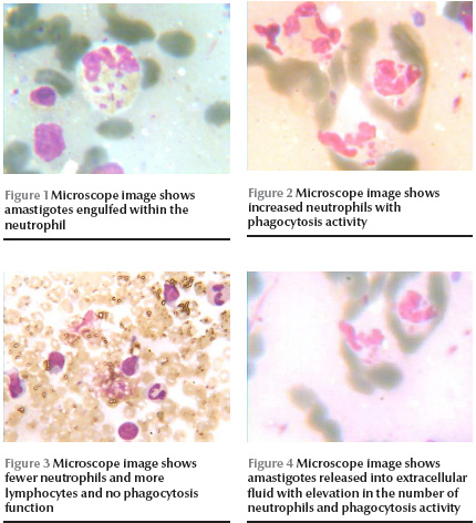
It appears from the sample microscope images that once the neutrophils became the predominant cell type in the field, their function in phagocytosis of the amastigotes becomes apparent (Figures 1 and 2). When the neutrophils were in lower concentrations, phagocytosis of amastigotes was not seen (Figure 3). The greater appearance of neutrophils when the amastigotes were present in the extracellular fluid, as shown in the cell counts, is evident in Figure 4.

Discussion
The cases in this study were referred with clinical signs of cutaneous leishmaniasis and no definite disease stage was determined for any of them. We can assume that none of the cases were at the very beginning of the disease process (as presented in other studies [2–4]) as studies indicate that no immediate clinical symptoms appear at the very early stage of the disease soon after the sand-fly transmits the parasite to the host [7,8]. When a patient presents clinically with a skin ulcer measuring 2–5 cm in diameter with a wet exudate and the pathological features show a dermal infiltrate mainly composed of macrophages filled with amastigotes, lymphocytes and plasma cells, then this is an early stage of the disease process. When the lesions clinically appear smaller in size, dry and nodular with dried exudates and the microscopic features show a decrease in the number of, or a disappearance of, the amastigotes and their macrophages, leaving a granulomatous infiltrate consisting of epithelioid cells, multinucleated giant cells, fewer lymphocytes and scanty plasma cells [7,8], then this is a later stage of the disease process.
In the microscope images, neutrophils are shown phagocytosing the released amastigotes in the extracellular fluid of infected lesions. Neutrophils are believed to be specialized in the phagocytosis process, while macrophages are considered less efficient than PMN neutrophils at killing foreign microorganisms, and the mechanism is not as well understood [9]. Accordingly, we can assume that those PMN neutrophils have a strong role to play in phagocytosis and destruction of amastigotes released after membrane rupture of the infected macrophages. Otherwise it is not possible to explain the disappearance of the amastigotes from the infected tissues after their release in huge numbers into the extracellular fluid later in the healing process of cutaneous leishmaniasis [7].
We can hypothesize that once the amastigotes are released from the ruptured phagocytes into the extracellular fluid in the late stage of the disease process, they are recognized by the neutrophils as foreign bodies and an acute-phase reaction is established. More neutrophils are attracted, as seen by an increase in the neutrophil concentration in the damaged area. The neutrophils start the process of phagocytosis of the released amastigotes and consequent limitation of disease.
In conclusion, this study has found that PMN neutrophil cells were at no time absent from the disease process of cutaneous leishmaniasis, in either the early or late stages. While previous studies have noted the role of PMN neutrophils in the early stages of the disease process, this study concentrated on their role in the later stages. The neutrophils seemed to be highly effective in phagocytosis of amastigotes released from ruptured phagocytes to the extracellular fluid. The PMN neutrophils seemed to establish their role by phagocytosis of the amastigotes as demonstrated by the disappearance of amastigotes when neutrophil counts drop.
References
- Daboul MW. Is the amastigote form of leishmania the only form found in humans infected with cutaneous leishmaniasis? Labmedicine, 2008, 39(1):38–41.
- Rousseau D et al. In vivo involvement of polymorphonuclear neutrophils in Leishmania infantum infection. BMC Microbiology, 2001, 1:17.
- Lima GM et al. The role of polymorphonuclear leukocytes in the resistance to cutaneous leishmaniasis. Immunology Letters, 1998, 64(2–3):145–151.
- Peters NC et al. In vivo imaging reveals an essential role for neutrophils in leishmaniasis transmitted by sand flies. Science, 2008, 321:970–974.
- Von Stebut E. Immunology of cutaneous leishmaniasis: the role of mast cells, phagocytes and dendritic cells for protective immunity. European Journal of Dermatology, 2007, 17(2):115–122.
- Launois P, Tacchini-Cottier F. Immune responses to leishmania infection [online report]. WHO Immunology Research and Training Centre, University of Lausanne, Department of Biochemistry (http://www.unil.ch/ib/page9491.html#1, accessed 1 July 2010).
- Hepburn NC. Cutaneous leishmaniasis: an overview. Journal of Postgraduate Medicine, 2003, 9:50–54.
- Sharquie KE et al. Evaluation and diagnosis of cutaneous leishmaniasis by direct smear, culture and histopathology. Saudi Medical Journal, 2002, 23(8):925–928.
- Sheehan C, ed. Clinical immunology: principles and laboratory diagnosis. Philadelphia, Lippincott–Raven, 1997.


 Volume 31, number 5 May 2025
 WHO Bulletin
 Pan American Journal of Public Health
 The WHO South-East Asia Journal of Public Health (WHO SEAJPH)