


Index Medicus for the Eastern Mediterranean Region
Eastern Mediterranean Health Journal
Select your language

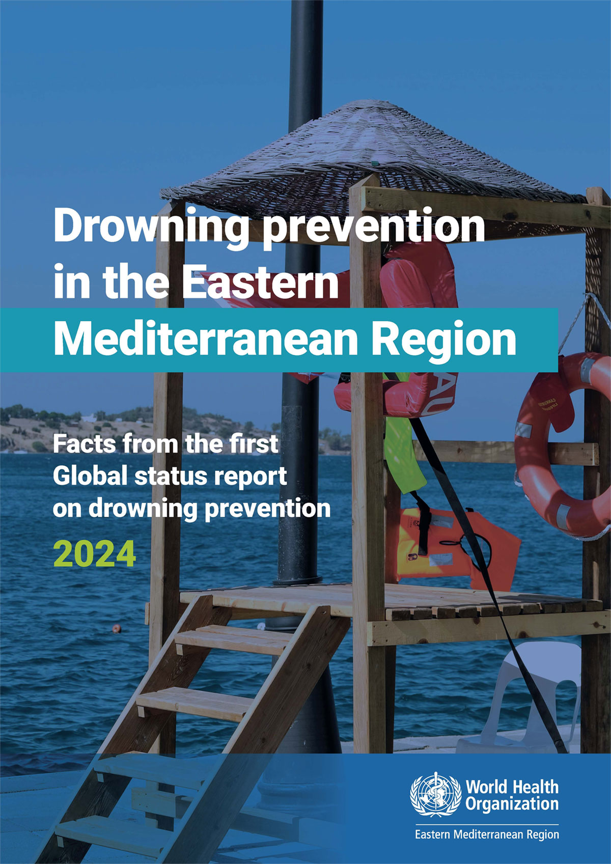
Drowning prevention in the Eastern Mediterranean Region. Facts from the first Global status report on drowning prevention, 2024 Globally, at least 3 million people have lost their lives to drowning over the past decade. An estimated 300 000 of these drowning deaths occurred in 2021 alone and 43% of these were children aged 14 years or younger. In 2021, drowning prevention became more prominent on the global agenda when the United Nations General Assembly adopted its first ever resolution on...
Bridging the divide. A guide to implementing the humanitarian-development-peace nexus for health (second edition) Despite growing consensus on the value of the humanitarian development and peace nexus (HDPNx) approach also known as the New Way of Working practical guidance on how to apply it effectively in the health sector has long been lacking. The HDPNx approach seeks to leverage the comparative advantages of different actors to improve efficiency and sustainability reduce duplication and...
Evidence-informed policy-making: a glossary of key terms A significant challenge within the ecosystem of evidence-informed policy-making is the absence of a shared vocabulary and consistent application of related terminology. Although various fields – including health, research, epidemiology and policy-making – offer definitions for certain terms, few are specifically tailored to evidence-informed policy-making, and none offer a fully comprehensive definition. As a result, there is...


WHO staff and visitors to the Regional Office are encouraged to visit the library and access a range of print materials and electronic resources available at the resource desk.
